Introduction
Newer inventions in techniques, equipment along with newer material has made endodontic surgery a preferred treatment modality in clinical situations where periapical pathology cannot be resolved by initial endodontic therapy or non surgical root canal treatment.1
Surgical procedure involves exposure of the root apex, root end resection, root end cavity preparation followed by insertion of root end filling material.2
Properties of a root end filling material play a very critical role in determining success of periradicular surgery. They seal apical region to avoid bacterial infiltration and their products from periradicular tissues to root canal system.3, 4 Gartner and Dorn explained the relation between clinical success and properties of an ideal rootend filling material, namely biocompatibility with the tissue fluids, dimensionally stablility in the presence of moisture, apical sealing abilitity and physical properties.5, 6
Throughout dental history wide varieties of rootend materials have been developed such as amalgam, glass ionomers, composite resins, zinc oxide eugenol cement, cavit, IRM, super EBA, and mineral trioxide aggregate. However, no single material has been able to satisfy all the requirements of an ideal root end filling material.7, 8, 9
Bioceramics are materials which include alumina, zirconia, bioactive glass, hydroxyapatite, calcium phosphates, glass ceramics. Commonly used Bioceramics in endodontics are Portland cement, MTA, Biodentine, calcium enriched mixture etc. Due to their similarity with biological hydroxyapatite they are excellent biocompatible materials.
Biodentine with active biosilicate technology is one such new calcium silicate based material with few properties superior to MTA like, has better consistency, handling properties and faster setting time.10
The development of ultrasonic have revolutionized root end therapy by improving the surgical procedure with better access to the root end in a limited working space resulting in better canal preparations, smaller osteotomy for surgical access, decrease number of dentinal tubules exposure and minimize leakage by avoiding giving bevelas compared to diamond points.11, 12
Current methods to evaluate the efficiency of degree of marginal adaptation are radioisotopes, scanning electron and confocal laser scanning microscopy, fluid filtration techniques.
In the present study scanning electron microscopy has been used to analyses marginal adaptation.
Materials and Methods
In this study, 56 freshly extracted human mandibular premolar teeth that were extracted for orthodontic and periodontal reasons were selected under protocol approved by the human research ethics committee. Teeth were initially disinfected by 5.25% sodium hypochlorite for 5 hours followed by storage in saline. Inclusion criteria was single rooted mandibular premolar teeth, radiographs were taken to rule out extra canals, calcifications, resorptions and root caries.
Crowns were decoronated above cemento-enamel junction to standardize the working length of specimens of about 14mm. pulp was extirpated, using nickel titanium rotary system ((Dentsply, Maillefer, Ballaigues, Switzerland) cleaning and shaping of canals was done, canals were then obturated using thermoplastic size dguttapercha and AH plus sealer.
Then the teeth were wrapped in wet gauze and placed in an incubator at 37°C for 24 hours for complete setting of the filling materials. Radiographs were taken to confirm the quality of obturation. Cavities were sealed with cavit.13
Apical three millimeters of roots were resected perpendicular to the long axis of the tooth with a high-speed handpiece and a diamond disc under constant water spray.
The samples were then randomly divided into two groups, group 1 and group 2 of 28 samples each. Group 1 and group 2 were further divided into subgroup A and subgroup B of 14 samples each. Root end preparation in subgroup A of both the groups were done with ultrasonics and subgroup B with diamond points of dimension 3x1mm.
Both the materials were mixed according to manufacturer’s instructions and cavities were filled using MTA carrier. In group I all the root end preparation were filled with MTA(angelus) and in group II with biodentine.
All the restored samples were allowed to set at room temperature for 24hours.
The apical portions of the roots were then sectioned to obtain 1mm thick traversal sections. The samples were examined under 100x and 500x magnification using a scanning electron microscope (SEM) to determine adaptations of the root end filling materials with the dentin in micron meters. The gap area was measured using software Image J.14
The values collected was analysed using two-way A nova and Tukeys multiple post hoc test.
Results
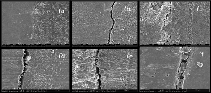
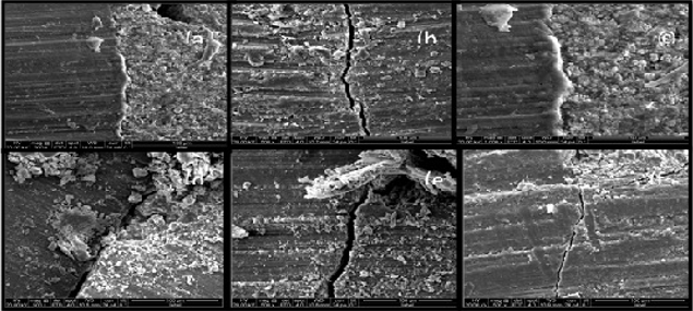
The scanning electron microscope of transverse sections of root end filled teeth showed marginal gaps at dentin -rooted filling interface (Table 1/Figure 1, Figure 2 ).
kolmosorov-smirnov test indicated data is normal and all variables follow a normal distribution therefore the parametric two-way A nova and Tukeys multiple post hoc procedures were applied.
Comparison of two groups (I, II) and two sub groups (A, B) with respect to marginal gaps by two-way ANOVA showed, between the main group there is significant difference seen (p= 0.0001). Lowest mean marginal gap of 0.46
Tukeys multiple post hoc procedures was carried out to compare the marginal gaps between Two Groups (I, II) and two sub groups (A, B) which revealed significant differences between all the groups(p<0.05).
Figure 1
SEM image under 500x magnification: MTA (Angelus) used as root end filling materials in which root end cavity was prepared by ultrasonicretrotip used (A-C) and root end preparation done with diamond point (D-F).

Figure 2
SEM image under 500x magnification:Biodentine used as root end filling material in which root end cavity was prepared by ultrasonic retrotrip used (A-C) and Root end preparation done with diamond points (D-F).

Table 1
Mean, SD, SE and coefficient of variation of marginal gaps in two groups (I, II) and two sub group (A,B)
Discussion
The aim of root end filling is to prevent movement of microorganisms and their byproducts from root canal into periapical tissues and vice versa. Properties of a root end filling material are very critical in determining success of periradicular surgery.5, 15
According to Gartner and Dorn an ideal material to seal root end cavities should; be biocompatible with the tissue fluids, dimensionally stable, the presence of moisture should not affect its sealing ability, be radioopaque to be recognized on the radiograph, prevent leakage of microorganisms and their by products into periradicular tissues.5
Numerous materials have been suggested as root end filling materials, In this study MTA (angelus) and Biodentineas root end filling materials were used as root end filling materials.
Mineral trioxide aggregate (MTA) was pioneered by Torabinejad at lomalinda university in 1993. Major constitiuents are tricalcium silicate, tricalcium aluminate, tricalciumoxide, silicate oxide, bismuth oxide, calcium carbonate. It has favorable properties suitable for an root end filling material such as excellent sealing ability, biocompatibility, good compressive strength (67Mpa), insoluble in fluids once set, radiopacity and antibacterial effect on some facultative bacteria (freshly mixed and 24 h set ) and C. albicans.1, 5
It shows the formation of calcium-phosphate precipitation at the interface. This interface layer reduces the risk of marginal percolation and gives promising long-term clinical success. Despite high clinical efficacy there are some issues which prevented clinicians to use it in many cases, major ones being, long setting time which might contribute to leakage, surface disintegration, loss of marginal adaptation and low compressive strength.12 Hence MTA angelus was introduced.
MTA (Angelus) was introduced in 2001 which contained 80% Portland cement and 20% bismuthoxide. In this restorative material, the calcium sulfate had been removed to reduce the setting time (17 minutes). MTA (Angelus) is compatible with the human body, has no mutagenic properties, does not cause apoptosis and also has antimicrobial properties and acceptable cytotoxicity, better handling properties and faster setting time.5, 15
Biodentine™ (Septodont, Saint- Maur-des-Fosses, France) it share mode of action as calcium hydroxide but does not have its drawbacks. powder mainly contains tricalcium and dicalciumsilicate, the principal component of Portland cement, as well as calcium carbonate. Zirconium dioxide serves as radiopacifier. The liquid consists of calcium chloride in aqueous solution with and mixture of polycarboxylate.5, 10
The liquid contains calcium chloride as an accelerator and a hydrosoluble polymer that serves as a water reducing agent. The setting period of the material is 9–12minutes. The material is characterized by the release of calcium when in solution. Tricalcium silicate based materials are also defined as a source of hydroxyapatite when they are in contact with synthetictissue fluid. The sealing ability of Biodentine is most likely through the formation of tags the calcium and silicon ion uptake into dentin leading to the formation of tag-like structures in Biodentine was higher than MTA(angelus), during setting of cement calcium phosphate is formed.5, 11
In the present study the marginal adaptation of both the filling materials are influenced by the root end preparation technique. In all the samples which were filled with either biodentineor
MTA (angelus), ultrasonically root end prepared samples showed less leakage values when compared to those prepared with diamond points which is similar to results found in previous studies. This can be attributed to the condition of cavity surface left after the preparation technique.4, 5
Cavities prepared with diamond points are left with greater amount of debris and smear layer in comparison to those prepared with ultrasonic tips. smear layer prevents complete contact between filling material and cavity walls hence explaining the greater leakage observed with both root end filling materials in cavities prepared with diamond points compared to ultrasonically prepared cavities. 15
