Introduction
Digital impressions represent innovative methods that enable dentists to construct virtual, computer-generated copy of hard and soft tissues of oral cavity, with use of lasers and other optical scanning machines. Digital method captures impression data with great accuracy, in minutes, without need for traditional impression resources that some patients find inopportune and messy. Patients consider digital impressions to be comfortable and easier method, in comparison with classical impression techniques. Impression information is then moved to computerized workstation that creates restorations, often without need for stone models.1
Space maintainer fabrication remains routine procedure for pediatric and general dentists. Space maintenance is typically recommended as an interceptive treatment to reduce complex orthodontic treatment at later age.2 Further, maintaining arch length becomes concern with loss of primary 2nd molars, unilateral loss of primary canines, or loss of 1st primary molars before eruption of permanent 1st molars. Most common method of obtaining an impression for space maintainer, an alginate impression with subsequent dental stone model, has disadvantages offending to distort over time as water evaporates from or absorbs into impression thereby causing inaccuracies in impression and subsequent stone casts. 3, 4 In this case series we will fabricate space maintainer and zirconia crown using digital impression thereby representing the systemic application of digital dentistry in pediatric patients.
Maintaining functional and aesthetic aspects, contributing to keep normal articulation and securing necessary space for eruption of permanent teeth are some of reasons why more primary teeth are treated by practitioners. These days, dentist is more interested toward treating and restoring primary teeth rather than extracting them. Despite new technologies used in order to improve dental materials in last decade, restoring primary teeth still contributes to difficult challenge in paediatric dentistry. When a child patient comes to clinic dentist must have knowledge, skills, materials and good behavioral management but these are not enough alone. Parental satisfaction is also necessary. 5
Behavioral issues of an apprehensive or uncooperative patient can be particularly problematic when clinician is trying to make conventional intraoral impression for appliance fabrication. In such cases, use of digital intraoral impressions would eliminate need for conventional alginate impression. Since impressions are considered an unpleasant experience by some children, switch to digital impression procedures may have long‐term positive impact on patient perceptions of dental procedures. In a study, measurements for orthodontic treatment planning were compared with dental stone and 3‐dimensionally printed models; no significant differences were found. 6
Once impression is captured, it can be either sent to laboratory, chairside system receives all information needed.7 Restoring deciduous teeth with crowns has many advantages: longevity, ease of application with less restorative microleakage.
The current case series describes the use of an intraoral scanner to make a digital impression for fabrication of a distal shoe space maintainer and fabrication of zirconia crown. This method reduced chair time in an uncooperative patient.
Case Report
Case 1
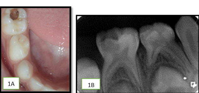
An 8-year-old boy came to clinic with decayed tooth and pain in mandibular back region. A medical and clinical history was taken along with the radiographic examination, which showed the presence of deep dentinal caries with pulpal exposure and extensive loss of tooth structure i.r.t right primary mandibular 2nd molar. Tooth was tender on percussion. (Figure 1a,b)
The tooth required a full-coverage restoration after endodontic treatment; both the child and the parent were highly concerned with the aesthetic appearance of the restoration. We decided to restore the primary mandibular right second molar with a paediatric zirconia crown using intraoral scanning and chairside milling.
We achieved local anaesthesia with NOIS (Nitrous Oxide Inhalational Sedation). We have modified our protocol here for the procedure exclusively for digital dentistry in paediatric patients; here we directly went on for tooth preparation, thereby utilizing the time taken by local anaesthetic to express its effect.
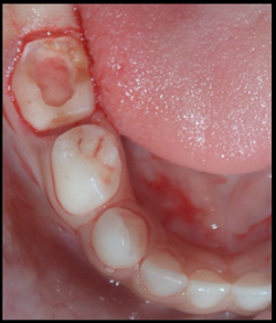
Scanning was done by intraoral scanner (Dentsply Sirona Omnicam) after minimal tooth preparation to capture a digital impression for further processing in CEREC software. [Figure 2]
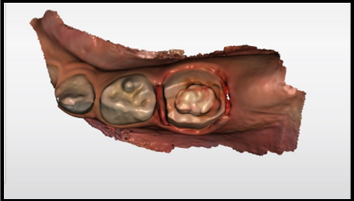
A Digital record of the segment with opposing arch was also recorded. Scanning provides easier, more intuitive, and precise 3D models in natural colour in less than 2 minutes. [Figure 3]
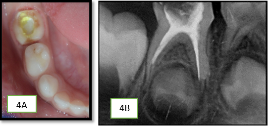
Now we perform Pulpectomy meanwhile designing and milling was done by supporter staff. [Figure 4a,b]
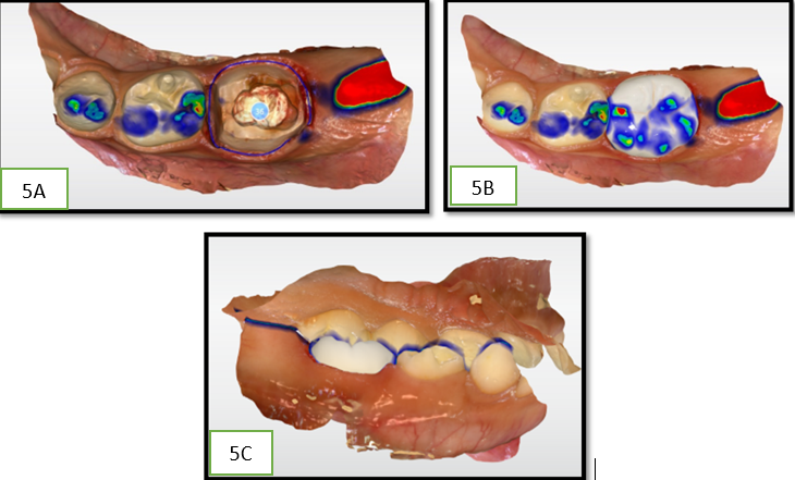
Designing was done in 5 minutes followed by milling in the primemill which took around 11minutes. [Figure 5A-C]
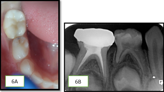
By the time pulpectomy was done we utilize time by preparing zirconia crown with chairside milling. Sintering was done in speedfire which provide adequate strength followed by glazing, done prior to cementation which makes surface more plaque resistant. Tooth and crown were cleaned of all blood residues. Glass ionomer cement (Fuji One PLUS, GC, Louvain, Belgium) was used for the cementation. [Figure 6A,B]
Excess cement was removed from interdental spaces and occlusion was checked. The patient was given post-operative instruction. [ Figure 7A,B]
In a matter of less than 45 minutes, complacent, enjoyable, and acceptable results were achieved. As shown in this case, we reconstituted not only the form and function but also the aesthetics with the help of digital scanner in a single sitting.
Case 2
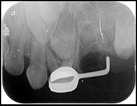
A 4.5-year-old child reported to the clinic with chief complaint of pain in the right upper back tooth region since10 days. OPG was taken which revealed radiolucency i.r.t primary maxillary 2nd molar. IOPA revealed grossly carious primary maxillary 2nd molar of right side (Figure 8 A,B)
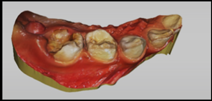
Looking at condition of tooth, extraction was planned with placement of willet’s appliance or distal shoe space maintainer, as to preserve arch length and maintain space for permanent successor tooth. Distal shoe space maintainer helps in prevention of mesial migration of erupting 1st molar successor tooth. Treatment was explained to parents and child. Scanning was done using CEREC Omnicam scanner before extraction on first appointment. (Figure 9)
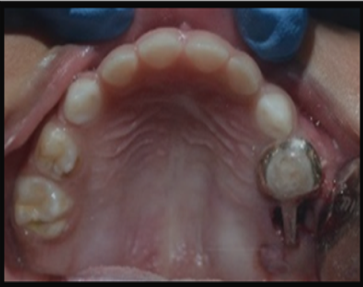
After 5 day of receiving the space maintainer from lab, patient was recalled. Post to the extraction, immediately distal shoe space maintainer was placed and cemented using GIC (Glass ionomer cement). (Figure 10) The intra-alveolar projection of the appliance was placed into socket to touch and guide vertical eruption guide path for unerupted permanent 2nd molar. (Figure 11)
Patient was given instructions and was recalled every 2 months to check for the condition of the appliance.
Discussion
Digital technology is transforming world, making produre easy for patient and dentist both. It makes visit for patient more efficient and pleasant. With 3D modeling and software such as CAD- CAM, dentist can plan restorative procedures easily with fewer errors. IOS, apart from obtaining impressions, detects obtained impressions suitable for modeling of restorations.8 Digital impressions contribute to precise registration of bite and help to skip various procedures that may cause distortions. 9 It becomes difficult to take impression with old methods but with these digital impression technique chances of errors is reduced with more precise impression which can be easily recorded.
Space maintainer fabrication is routine procedure thesedays for pediatric and general dentists. Space maintenance is typically recommended as an interceptive treatment to reduce complex orthodontic treatment at later age. 10
Further, maintaining arch length becomes concern with loss of primary 2nd molars, unilateral loss of primary canines, or loss of 1st primary molars before eruption of permanent 1st molars. Most common method of obtaining an impression for space maintainer, an alginate impression with subsequent dental stone model, has disadvantages offending to distort over time as water evaporates from or absorbs into impression thereby causing inaccuracies in impression and subsequent stone casts & time lapse for model or impression to reach lab and other state or city. 11, 12
Complete digital workflow through chairside systems allows indirect restorations to be carried out in same clinical session in which preparations are made. This prevents temporization, inflammation of tissues, greater comfort for patient and a rapid workflow for dentist. 13
Advantages of digital impression include simplicity, elimination of “dirty” cabinet and patient discomfort. There is no issues of incorporating air bubbles in impression. However, close attention should be directed towards not acquiring artifacts due to saliva, gum margins, and sites in oral cavity. There is no need for storage space or purchase of spoons and impression materials. Risk of contamination is reduced, and disinfection needed by classical impressions is not a must for digital impression technique. 14
Aesthetic treatment of severely decayed primary teeth is greatest challenges in pediatric dentistry. Use of aesthetic restoration has become an important aspect in pediatric dentistry. Over years, numerous techniques for restoring primary teeth have been attempted. This case reports describes a single-visit chairside fabrication of zirconia crown on a primary molar and in other case fabrication of space maintainer is represented with the help of digital workflow. To limit chairside time, CAD/CAM technology could be used in cases where crown is needed in primary teeth. With the help of the digital design software technology, dentist can provide better minimally invasive crowns without taking traditional impression techniques especially in non-competent child patients.
Conclusion
The CAD/CAM technologies are increasingly being used in aesthetic dentistry in both adults and children. Digital impression technique can be easily applied to restore children’s dentition with contemporary metal-free ceramic constructions in order to provide better aesthetic and functional crowns and space maintainers in duration short time in pediatric dentistry.

