- Visibility 401 Views
- Downloads 23 Downloads
- DOI 10.18231/j.ijce.2024.044
-
CrossMark
- Citation
Restoring with flowables: An injection moulding technique – A report of two cases
Introduction
In aesthetic dentistry, the primary focus is on minimally invasive treatments that preserve as much dental structure as possible while achieving desirable aesthetic results. Ceramic veneers are often regarded as the gold standard for these procedures due to their excellent mechanical properties and long-lasting aesthetics. However, recent advancements in composite resin have led to a rise in the popularity of composite veneers. These veneers present several benefits: they bond and repair easily, have a higher flexural modulus, are more cost-effective, and produce less wear on opposing teeth.[1]
Creating direct or indirect final restorations is a meticulous and time-consuming process that requires specific skills and careful attention to detail from the clinician. Traditional direct anterior restorations are particularly difficult due to the intricate layering technique involved, which restricts their optimal use to only a few restorations at a time.
When choosing composite resin, clinicians should take into account factors such as durability, aesthetic properties, and ease of handling. Composite resins have seen significant advancements in both optical and mechanical properties, improving their versatility despite some limitations. Flowable composite resins, introduced in late 1996, represent a variation of conventional composites with a lower filler content of 37%-53% by volume, compared to the 50%-70% by volume found in conventional mini-filled hybrid composite resins.[2]
The integration of nanotechnology into flowable composite resins has led to materials that combine the mechanical properties, wear resistance, strength, enhanced polishability, excellent polish retention, and translucency of conventional resin composites with the elasticity, adaptability, and favourable handling characteristics of flowable composites. This advancement also reduces polymerization shrinkage by nearly 20%.[3]
The injectable resin composite technique offers an innovative approach that merges both indirect and direct methods to accurately and reliably transform a diagnostic wax-up into composite restorations. Traditionally, the free-hand direct technique has been utilized, necessitating a highly skilled clinician and frequently leading to unpredictable results. To overcome these challenges, various clinicians have introduced restorative concepts that simplify the workflow, making it more efficient and less stressful while providing more consistent outcomes.
One recommended approach for anterior extended restorations is the injection moulding technique. This method starts with the creation of a transparent silicone matrix based on a diagnostic wax-up. The silicone matrix extends to adjacent unrestored teeth, ensuring precise fit and accurate positioning during the restorative process. The clinical applications of this technique are varied and include the emergency repair of fractured teeth and restorations, the fabrication of provisional restorations (as noted by Terry in 2012), and the creation of transitional composite restorations such as Class III, Class IV, and veneers. Additionally, it is used in pediatric composite crowns, resurfacing occlusal wear on posterior composite restorations, establishing the correct incisal edge length before aesthetic crown lengthening, and creating composite prototypes for copy milling.
This comprehensive approach not only improves the precision and predictability of composite restorations but also broadens the range of possible applications, offering a versatile solution for various clinical situations. By utilizing the benefits of both direct and indirect techniques, the injectable resin composite method signifies a major advancement in restorative dentistry.[4], [5]
This case report discusses the treatment plan for three patients using this composite resin injection technique to improve their compromised anterior aesthetics.
Case Report
Case 1
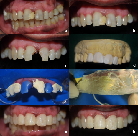
A 47-year-old male patient presented with a primary concern of brownish discolourations in the upper front region, which affected the appearance of his smile. Upon examination, multiple fractured restorations and secondary cavities were identified from the upper left to the right lateral incisors. Additionally, the patient had a diastema and maxillary protrusion ([Figure 1] a,b).
With the patient's consent, the injectable composite resin technique was chosen, as it met his needs for a quick, minimally invasive, and cost-effective solution. During the initial appointment, essential photographs were captured to aid in the smile design phase. The fractured restorations and cavities were removed, and putty and light-body polyvinyl siloxane impressions (GC Flexceed, GC India Dental Pvt., India) were used to create a diagnostic cast. A wax mock-up was then prepared, extending from the upper left canine to the upper right canine ([Figure 1] c,d).
At the second appointment, temporary veneers made of acrylic resin were created based on the wax mock-up to evaluate aesthetic and occlusal parameters before the final restorations. Factors such as tooth length and shape, lateral and protrusive movements, maximum intercuspation, and veneer thickness were assessed. Once these parameters were deemed satisfactory by both the dentist and the patient, the fabrication of the definitive veneers using the injectable composite resin technique was initiated.
To produce a silicone template for the diagnostic wax mock-up, transparent silicone (Exaclear, GC Corporation, Tokyo, Japan) was placed in a solid metal impression tray. The fit of the template in the patient’s mouth was verified. Injection holes for the flowable composite were drilled using fine chamfer diamond burs, aligning them with the incisal edges. Rubber dam split isolation was employed, with each tooth designated for composite injection isolated from adjacent teeth using a 0.075 mm thick strip of polytetrafluoroethylene.
The process began with acid etching using 37% phosphoric acid (D-tech XT Etch, India) for 15 seconds, followed by rinsing and applying adhesive (G-Premio Bond, GC Corporation, Tokyo, Japan). After curing for 20 seconds, the silicone template was positioned, and a flowable composite in shade A3 (G- ænial Universal Injectable, GC Corporation, Tokyo, Japan) was injected through the incisal edge hole, filling the space from the cervical to the incisal area between the tooth and silicone template. Once filled, the material was light-cured with an LED curing light for 40 seconds ([Figure 1] e,f).
After template removal, any excess material was excised using a 12D scalpel blade and dental probe. Subsequently, definitive light curing of the vestibular surfaces was performed for 20 seconds. This process was repeated for each tooth, and the finishing of all restorations was carried out using a fine diamond bur from Mani Inc., Japan. Interproximal polishing was done with fine and extra-fine interproximal strips from Shofu Inc., Kyoto, Japan. The labial surfaces of each tooth were refined with Sof-Lex Extra Thin contouring and polishing discs from 3M ESPE, St. Paul, MN, USA, and final polishing was completed using rubber polishers and diamond polishing paste from the Prisma Gloss composite polishing paste system by Dentsply DeTrey GmbH, Germany, to prevent plaque accumulation and staining ([Figure 1] g,h).
Case 2
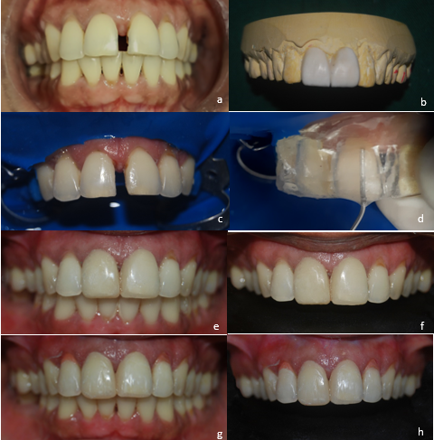
A 32-year-old female patient presented with a primary concern of a midline diastema that negatively impacted her smile. Further examination revealed caries affecting the upper right and left central and lateral incisors. Additionally, the patient exhibited Class 1 gingival recession according to Miller's classification (1985) in the upper right and left lateral incisors. The treatment approach was similar to that of Case 1. For this patient, the selected shade was A2. After completing the final finishing and polishing of the restorations, the gingival recession was addressed using a gingival composite in shade G-DP (Beautifil II, Shofu Inc., Kyoto, Japan) ([Figure 2]).
Case 3
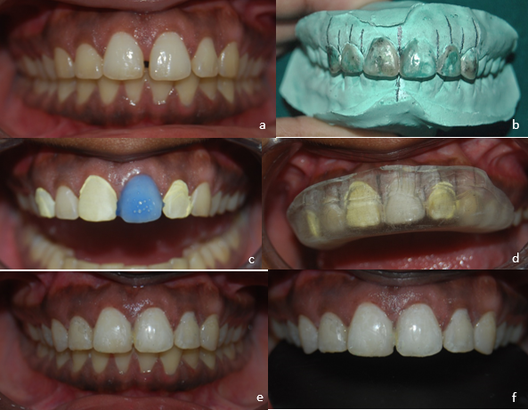
A 26-year-old female patient presented with a primary concern of a diastema in the upper front region, which was negatively impacting her smile. Upon closer examination, a diastema extending from the upper right to left canine was observed, along with attrition on both quadrant canines. The treatment procedure closely mirrored that of Case 1, with the A2 shade being utilized ([Figure 3]).
Final photographs were taken immediately for all patients, and they expressed satisfaction with the treatment results regarding both function and aesthetics.


Discussion
A range of methods has been proposed to ensure the precise replication of anatomical features in aesthetic composite restorations for anterior teeth. These techniques include the use of silicone indexes to capture the palatal surface of restorations and the employment of transparent silicone indexes to simultaneously replicate the entire surface anatomy, as demonstrated in this case report.
This paper presents a straightforward technique that can be easily adapted to recreate anatomical morphology, restore functionality, and enhance the natural aesthetics of permanent teeth. By integrating these methods into a systematic workflow, clinicians can achieve consistent and predictable results in the restoration of anterior teeth, ultimately contributing to improved patient satisfaction and treatment outcomes.
Flowable composite resin is often favoured for use with transparent silicone indexes due to its ability to accurately replicate the prepared wax-up intraorally with minimal effort. Unlike conventional composite materials, which may require significant external pressure on the index to achieve a precise reproduction of tooth morphology, flowable composite resin provides a more straightforward and reliable means of achieving accurate replication within the index.[6] During a 24-month observation period, no significant differences in clinical outcomes were noted between the use of a nanohybrid composite resin and a flowable composite for restoring non-carious cervical lesions.[7]
In anterior restorations, the chosen material must not only accurately replicate the natural tooth colour but also maintain this colour over time. Achieving an optimally finished restoration with a smooth surface is crucial, as this helps prevent plaque accumulation and resist staining. While conventional flowable resin composites offer advantages such as excellent adaptation to pulpal margins and ease of direct cavity filling using small gauge dispensers, they often suffer from lower filler particle content and higher organic content. Consequently, these materials display reduced mechanical properties, increased susceptibility to wear, diminished polishability, and compromised colour stability.
To address these limitations, manufacturers have developed new formulations of flowable resin composites with a higher filler content, typically around 69 wt%, which surpasses that of conventional flowable materials. Known as "highly filled flowable composites" or "injectable composites," these advanced materials aim to streamline the filling procedure, thereby reducing chairside time while providing improved mechanical and aesthetic properties compared to their predecessors. This innovative approach, as illustrated in the present clinical report, represents a significant advancement in anterior restoration techniques, promising better outcomes and enhanced patient satisfaction. [8], [9]
Modern highly filled flowable composites, featuring nanofillers smaller than 200 nm, now exhibit enhanced mechanical properties and improved aesthetics. Recent studies report no significant statistical or clinical differences in outcomes between these flowable composites and traditional composites. In a study by Kitasako et al., the clinical evaluation of conventional and highly filled flowable composites in direct restorations showed favourable results, with most receiving clinically acceptable ratings after 36 months. Notably, the highly filled flowable composite demonstrated low volume loss and produced a significantly smoother surface following wear tests compared to other flowable and conventional composites. Although this was tested in posterior direct restorations, the fine filler distribution of the highly filled flowable composite contributed to wear resistance on par with that of conventional composite materials.[10], [11]
Concerns about wear resistance have historically been linked to older composite restorative materials, particularly in direct posterior restorations. However, a recent two-body wear study demonstrated that the highly filled flowable composite (G-aenial Universal Flo) utilized in the current clinical assessment exhibited minimal volume loss, comparable to that of amalgam. Furthermore, it produced a significantly smoother surface after the wear test when compared to other flowable and conventional composites, such as GrandioSO Flow and GrandioSO Heavy Flow from VOCO (Cuxhaven, Germany), and Filtek Supreme XTE from 3M ESPE Dental Products (St. Paul, MN, USA).[12]
Recent research on highly filled flowable composites has demonstrated excellent surface characteristics following toothbrush abrasion, achieving a surface polish comparable to or even surpassing that of paste composites. However, these clinical studies focused on short-term follow-ups, with long-term outcomes yet to be explored.[13], [14]
One notable limitation of this case report is the absence of long-term follow-up data of at least 12-24 months regarding the durability of the restorations. Nevertheless, future clinical applications of this innovative technique using advanced flowable materials could provide clinicians and technicians with alternative strategies for managing various clinical situations. This would enable them to deliver improved and more reliable dental care to their patients.
Conclusion
The application of the injectable technique in this study was essential for achieving precise anatomical replication of the diagnostic wax-up. This approach significantly enhanced marginal accuracy and resulted in immediate outcomes that were both highly aesthetic and functional. Additionally, the workflow associated with this technique was straightforward and manageable, particularly when carefully planned and executed. Successful functional outcomes depend on meticulous planning and the preparation of a comprehensive wax-up, which serves as a blueprint for the subsequent intraoral restorations. By utilizing a transparent index, clinicians can accurately translate the intricate details of the wax-up into the final restorations. This method not only streamlines the process but also ensures consistency and predictability in achieving the desired treatment outcomes.
Conflict of Interest
None
Source of Funding
None.
References
- J Cortés-Bretón Brinkmann, MI Albanchez-González, DML Peña, IG Gil, MJS García, JP Rico. Improvement of aesthetics in a patient with tetracycline stains using the injectable composite resin technique: a case report with 24-month follow-up. Br Dent J 2020. [Google Scholar]
- C Coachman, L De Arbeloa, G Mahn, TA Sulaiman, E Mahn. An Improved Direct Injection Technique With Flowable Composites. A Digital Workflow Case Report. Oper Dent 2020. [Google Scholar]
- OO Shaalan, E Abou-Auf, AF El Zoghby. Clinical evaluation of flowable resin composite versus conventional resin composite in carious and noncarious lesions: Systematic review and meta-analysis. J Conserv Dent 2017. [Google Scholar]
- DA Terry, JM Powers. A predictable resin composite injection technique, Part I. Dent Today 2014. [Google Scholar]
- V Kouri, D Moldovani, E Papazoglou. Accuracy of Direct Composite Veneers via Injectable Resin Composite and Silicone Matrices in Comparison to Diagnostic Wax-Up. J Funct Biomater 2023. [Google Scholar] [Crossref]
- D Geštakovski. The injectable composite resin technique: minimally invasive reconstruction of esthetics and function. Clinical case report with 2-year follow-up. Quintessence Int 2019. [Google Scholar]
- E Karaman, AR Yazici, G Ozgunaltay, B Dayangac. Clinical evaluation of a nanohybrid and a flowable resin composite in non-carious cervical lesions: 24-month results. J Adhes Dent 2012. [Google Scholar]
- NRY Gia, CS Sampaio, C Higashi, A Sakamoto, R Hirata. The injectable resin composite restorative technique: A case report. J Esthet Restor Dent 2020. [Google Scholar]
- DA Terry, JO Burgess, JM Powers, MB Blatz. A Novel Concept for Developing a Precise and Predictable Post and Core Complex Using the Injectable Resin Technique. Compend Contin Educ Dent 2024. [Google Scholar]
- Y Kitasako, A Sadr, M F Burrow, J Tagami. Thirty-six month clinical evaluation of a highly filled flowable composite for direct posterior restorations. Aust Dent J 2016. [Google Scholar]
- K Shinkai, Y Taira, S Suzuki, S Kawashima, M Suzuki. Effect of filler size and filler loading on wear of experimental flowable resin composites. J Appl Oral Sci 2018. [Google Scholar] [Crossref]
- D Lazaridou, R Belli, A Petschelt, U Lohbauer. Are resin composites suitable replacements for amalgam? A study of two-body wear. Clin Oral Investig 2014. [Google Scholar]
- G Lai, L Zhao, J Wang, KH Kunzelmann. Surface properties and color stability of dental flowable composites influenced by simulated toothbrushing. Dent Mater J 2018. [Google Scholar]
- RL Wang, CY Yuan, YX Pan, FC Tian, ZH Wang, XY Wang. Surface roughness and gloss of novel flowable composites after polishing and simulated brushing wear. Zhonghua Kou Qiang Yi Xue Za Zhi 2017. [Google Scholar]
